

Real-Time Ultrasound
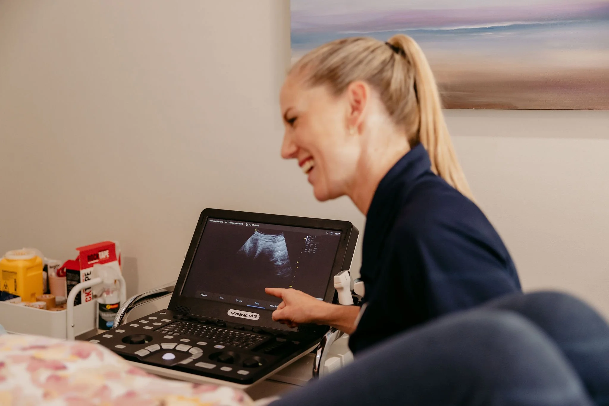
Real-time ultrasound gives your physio a live, on-screen view of your muscles, joints and soft tissues during assessment and treatment. It's a safe, non-invasive tool that helps us diagnose more accurately, guide treatment decisions and show you exactly what's going on — so you're never left guessing.

How Is Ultrasound Used in Physiotherapy?
Your physio uses a small handheld probe on the skin to produce a live image of the area being assessed.
It's commonly used to check how well deep stabilising muscles are activating (like your pelvic floor or deep core), to assess soft tissue injuries, and to monitor healing progress after surgery. It's painless, takes just a few minutes, and gives both you and your physio real-time visual feedback that makes treatment more targeted and effective..

Who Is It Most Useful For?
Real-time ultrasound is particularly valuable for women's health patients working on pelvic floor recovery, post-surgical patients who need to retrain deep muscle activation, and anyone dealing with an injury where understanding exactly what's happening beneath the surface makes a real difference to the treatment approach.
It's also a powerful motivator — being able to see your muscles working on screen helps you connect with the exercises and understand why they matter.